
23 Embryology of the Urinary System
EMBRYOLOGY OF THE URINARY SYSTEM
Learning Objectives
- Describe the three generations of the kidney, as well as the formation of the ureter and bladder from the intermediate mesoderm.
- Describe the ascent of the kidney from its original sacral location to the lumbar region and how congenital renal anomalies may appear.
- Understand how the urorectal septum partitions the cloaca of the hindgut into a primitive urogenital sinus, giving rise to the bladder and urethra, and a posterior rectum.
Lecture notes
Larsen, W.J. Human Embryology 4th Edition, Chapter 15, pp. 479-541
We also recommend reviewing the tutorial on the Urogenital System developed by the University of North Carolina at:
http://www.med.unc.edu/embryo_images/unit-genital/genital_htms/genitaltoc.htm
Overview of Development
The urinary and genital system are intimately associated in development. They both derive from a common mesodermal ridge (the intermediate mesoderm), along the posterior wall of the abdominal cavity.
The intermediate mesoderm gives rise to three successive nephric structures: 1) pronephros – primitive kidney that develops in the cervical region, 2) mesonephros – successor of the pronephros, develops in the thoracic and lumbar regions, and 3) metanephros – definitive kidney.
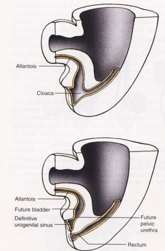
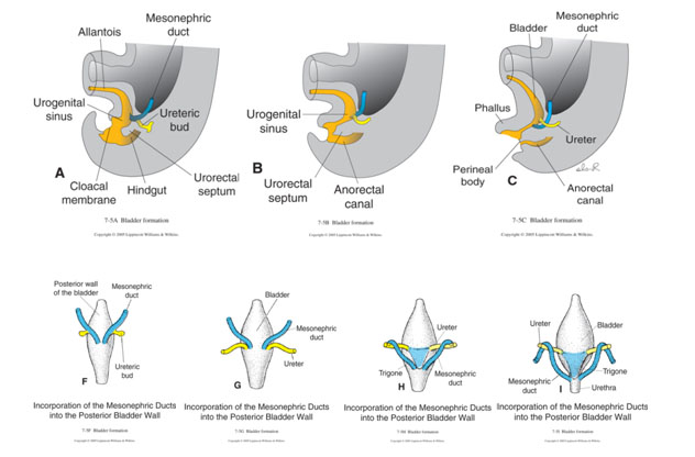
The cloaca is partitioned into a dorsal anorectal canal and a ventral urogenital sinus. The ventral urogenital sinus is continuous with the allantois, which projects toward the umbilical cord. The expanded superior portion of the urogenital sinus becomes the bladder, and its inferior portion gives rise to the pelvic and penile urethra (in males) and pelvic urethra and vestibule of the vagina (in females).
|
©Larsen’s Human Embriology 4th Ed. |
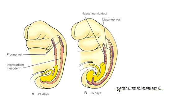
The pronephroi
- Develop early in the 4th week at the cervical level.
- Give rise to mesonephric (Wolffian) duct.
- Cease developing by day 24-25, never functional.
- Induce the development of the next stage of the kidney: mesonephric kidney.
- Mesonephric duct induces evagination of ureteric bud in both sexes, and disappears in females, but becomes ductus deferens in males.
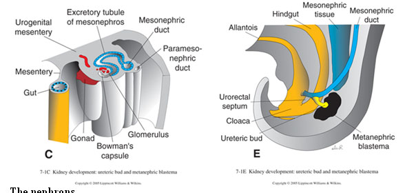
The mesonephroi
|
©Larsen’s Human Embriology 4th Ed. |
The metanephroi
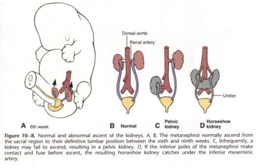
- Formation of the definitive kidney begins during the 5th week, with the induction and formation of a pair of ureteric buds within the intermediate mesoderm of the sacral region.
- Each ureteric bud sprouts from the distal mesonephric duct, penetrates the metanephric blastema, and begins bifurcation. Each tip (ampulla) acquires a cap-like aggregate, giving the metanephros a lobulated appearance.
- The ureters and collecting duct system differentiate from the ureteric bud. The nephrons (urine-forming units) differentiate from the metanephric blastema. The differentiation of each of these primordials depends on inductive signals from one another. The metanephric mesenchyme induces the ureteric bud to grow and branch to form the collecting ducts and tubules, whereas the tips of the ureteric buds induce the mesenchyme to condense and convert into an epithelial vesicle. If the ureteric bud is abnormal or missing, the kidney does not develop.
- In the mature kidney, urine produced by the nephrons flows through a collecting system consisting of collecting tubules, collecting ducts, minor calyces, major calyces, the renal pelvis, and the ureter, all products of the ureteric bud.
- Some parts of the kidney are formed by further branching, while other parts are formed by the coalescence of branches:
|
Kidney differentiation |
|
|
Kidney structure |
Formed by… |
|
Renal pelvis |
Expansion of the first 2 branches of the ureteric bud |
|
Major calyces |
The next 4 generations of branching |
|
Minor calyces |
Coalescence of the next 4 generations |
|
Collecting ducts |
An additional 11 generations of branching |
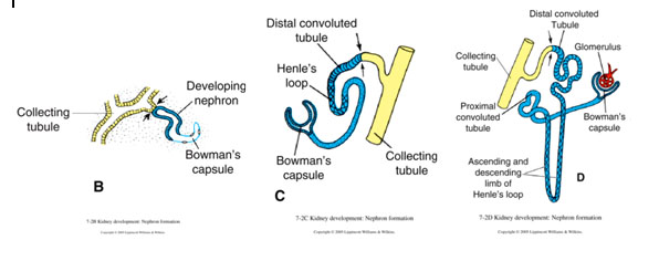
The nephrons
- Each nephron originates as an epithelial vesicle within the blastemic cap surrounding the ampulla of a collecting tubule (metanephric tissue).
- First, the nephric vesicle develops into a comma-shaped structure and then forms an S-shaped tubule, which fuses with the ureteric duct, and eventually the two lumina become continuous forming the uriniferous tubule.
- The renal corpuscle segment of the nephric tubule forms the outer layer of Bowman’s capsule. The lengthening nephric tubule forms the remaining elements of the nephron: the proximal convoluted tubule, descending and ascending limbs of the loop of Henle, and distal convoluted tubule. The medulla of the kidney also begins to take shape.
- The kidney is divided into an inner medulla and an outer cortex. The cortex contains the nephrons, whereas the medulla contains colleting ducts and loops of Henle.
- Nephrogenesis is complete by birth.
Development of urinary tract
The remainder of the urinary tract is derived from hindgut endoderm. The cloacal region is partitioned by the urorectal septum into a ventral urogenital sinus and a dorsal anorectal canal. The primitive urogenital sinus has 3 parts:
- The presumptive bladder cranially, continuous with the allantois.
- An intermediate narrowing that will become the pelvic urethra.
- A phallic segment beneath the genital tubercle. In males, the pelvic urethra becomes the membranous and prostatic urethra, and the phallic segment contributes to the penile urethra. In females, the pelvic urethra becomes the membranous urethra, and the phallic segment contributes to the vestibule of the vagina.
Distal portions of the mesonephric ducts and attached ureteric buds (future ureters) become incorporated into the posterior wall of the bladder, causing the mouths of the narrow part of the mesonephric ducts to migrate caudally until they open into the pelvic urethra, just below the neck of the bladder. The triangular area on the posteroinferior wall of the bladder is called the trigone of the bladder. The mesodermal tissue of the trigone is later overgrown by endoderm from the surrounding bladder wall, but it remains visible in the adult. The mesoderm associated with the bladder wall forms the smooth muscle of the bladder.
Anomalies of urinary development
- The kidneys can develop multiple renal vessels. These can occur as the kidney ascends and new vessels form but old vessels do not degenerate.
- There can be duplication of the ureter (bifid ureter). This occurs when the ureteric bud branches before it reaches the metanephrogenic tissue.
- The kidney can also form abnormally into a horseshoe kidney. In this condition the kidney is found in the region of the abdomen below the inferior mesenteric artery.
- Rarely a kidney fails to ascend, remaining as a pelvic kidney.
DEVELOPMENT OF THE URINARY SYSTEM – SUMMARY
The kidney is located on the posterior abdominal wall at the thoraco-lumbar vertebral level. The ureters travel inferiorly through the abdomen and cross the iliac arteries to enter the pelvis. During their course the ureters are retroperitoneal. As they course through the pelvis they enter the bladder posteriorly.
- Intermediate mesoderm gives rise to:
- Adrenal cortex
- Nephrogenic cord
- Pronephros(transient)
- Mesonephros (forms temporary kidney)
- Mesonephric duct (gives off ureteric bud in both sexes, disappears in females but becomes ductus deferens in males)
- Metanephros (definitive kidney)
- Paramesonephric duct (forms the uterine tube, uterus, and vagina, disappears in males)
- Gonadal ridge (gives rise to tissues of the gonad)
- Urogenital Sinus:
- Anterior part of the hindgut separated by growth of the urorectal septum from the rectum behind
- Becomes bladder and female urethra and forms prostatic and membranous parts of urethra in male
- Major Events and Topographic Changes:
- Division of cloaca by urorectal septum
- Delineation of intermediate mesoderm into mesonephros, gonadal ridge, and tubal ridge containing mesonephric (Wolffian) duct and paramesonephric (Müllerian) duct
- Growth of tubal ridges with their contained ducts caudally and medially to enter urogenital sinus dorsally
- Formation of ureteric bud from mesonephric duct and separation of opening into bladder of mesonephric and ureteric orifices
- Delineation of bladder and urethral components of urogenital sinus
- Development of the Definitive Kidney
- Branching of ureteric bud
- Formation of metanephrogenic caps as successive branching occurs
- Differentiation of metanephrogenic caps into glomeruli and nephrons and establishment of continuity of these with ureteric branches
- Absorption of proximal branches of ureteric buds to give main collecting ducts opening on renal papillae
- Major congenital anomalies of kidney and ureter
- Multiple renal vessels
- Duplication of ureter
- Horseshoe kidney
- Pelvic kidney
Embryology of the Urinary System quiz click here